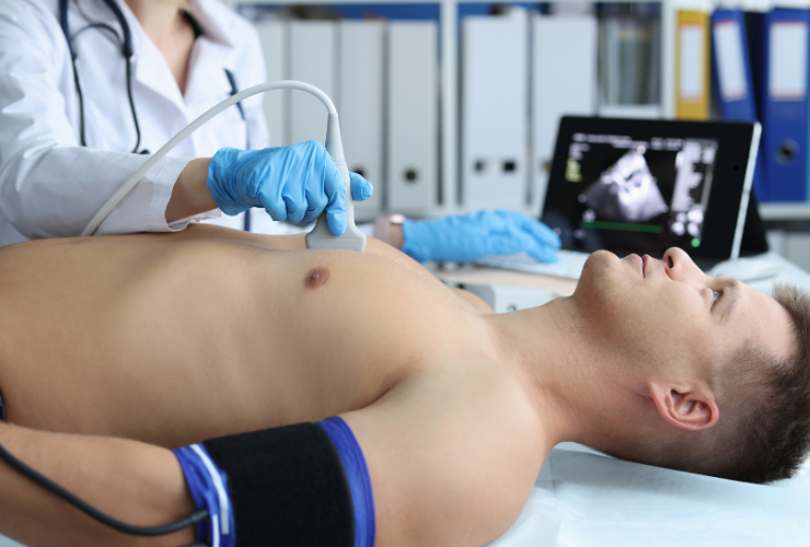


Welcome to Akshar Imaging Centre's dedicated page for 2D Echo (Two-Dimensional Echocardiography) testing. This non-invasive procedure plays a crucial role in evaluating the structure and function of your heart, aiding in the early detection and management of various cardiac conditions.
2D Echo uses ultrasound technology to create detailed images of your heart in real-time. These images help our specialists assess the heart's chambers, valves, and blood flow patterns, providing valuable insights into your cardiac health. Unlike other imaging techniques, such as MRI or CT scans, 2D Echo offers a dynamic view of your heart's movements and functions.
A 2D Echo test is recommended to diagnose and monitor a range of heart conditions, including:
Early detection through 2D Echo can significantly impact treatment outcomes by allowing timely intervention and management of heart conditions before they progress.
Preparing for your 2D Echo test is straightforward
During the procedure:
After your 2D Echo test:
Early detection through routine 2D Echo screenings offers several benefits:
Yes, 2D Echo is considered safe and non-invasive, using harmless ultrasound waves to create images of your heart.
No, the procedure is painless and generally well-tolerated by patients.
Unless instructed otherwise, you can eat and drink normally before your 2D Echo test.
The 2D Echo test is recommended in several scenarios, including diagnosing heart valve problems, assessing congenital heart defects, evaluating heart function post-heart attack, and monitoring overall cardiac health.
The 2D Echo test is required to provide detailed images of the heart's structure and function. It helps diagnose various heart conditions early, allowing for timely medical intervention and personalized treatment plans.
2D Echo Doppler combines traditional 2D Echo imaging with Doppler ultrasound to measure blood flow through the heart's chambers and blood vessels. It assesses the direction and speed of blood flow, providing additional insights into heart function and detecting abnormalities like valve regurgitation or stenosis.
A 2D Echo stress test, also known as a stress echocardiography, evaluates heart function during physical stress, typically induced by exercise or medication. It helps diagnose coronary artery disease and assess heart function under exertion.
During a 2D Echo cardiography, a transducer is placed on the chest to emit ultrasound waves. These waves create real-time images of the heart, showing its size, shape, and motion. The procedure is painless and allows cardiologists to assess heart health comprehensively.
No, pneumonia typically cannot be detected by a 2D Echo test. This test focuses on evaluating the structure and function of the heart, specifically its chambers, valves, and blood flow patterns. Diagnosis of pneumonia usually requires clinical evaluation, chest X-rays, and sometimes additional tests like blood tests or CT scans.
At Akshar Imaging Centre, we prioritize your cardiac health. Regular 2D Echo screenings are vital for maintaining heart wellness and detecting potential issues early. Schedule your appointment today to take proactive steps towards a healthier heart.